SUMMARY
High resolution colorimetric mapping of the Turin Shroud, by means of digital acquisition with a solid state camera, is proposed with the aim of obtaining a database for future accurate analysis. For example, more information about the mechanism of image formation on the cloth may be obtained.
This obviously implies proper calibration of the acquisition systems with uncertainty evaluation (done before, during and after mapping), considering some metrological problems such as the spatial and temporal stability of acquired signals, image distortions, dark currents and defective pixel effects.
1) INTRODUCTION
Many multidisciplinary studies carried out on the Turin Shroud [1], [2], [3], [4], [5], [6], [7], indicate that the probability that the image derives from a hypothetical craftsman trying to make an image on the cloth is extremely low.
Many authors have attempted to show that the figure on the Shroud does not represent Jesus Christ's image. For example, in 1988 [8] the medieval age of the linen was demonstrated, although bias errors due to fire of 1532, carboxidation of cellulose, C14 biofractionation in the linen plant, the presence of bioplastic coating [9], and the poor representativity of the Shroud samples, which were cut from a corner probably containing darns made in medieval times, were not taken into account.
In 1898, Secondo Pia made the first photographic image of the Man of the Shroud, developing a negative plate of the Turin Shroud. A century later, during the exhibition planned for next year, the first high resolution colorimetric mapping of the whole cloth may be achieved at various resolution levels. Digital mapping, useful for future studies, is proposed for the following aims:
- I) Providing a data base for future studies open to all scientists.
-
II) High resolution digital colorimetry at pixel level for quantitative analysis of the radiation source causing the image on the Shroud.
Quantitative analysis provides more information about radiation characteristics, for example, correlation between emitted radiation and human body mass is possible. Areas with higher radiation intensity may be investigated and correlated to relative parts of the anatomy.
Jackson et al. [7] observed that only the surface fibrils of the image appear on the Shroud to be discolored, and calculated the time necessary for an electromagnetic source to cause this effect. This time "must be of the order of hundredths of a second, a time which would pose considerable technical difficulties for a hypothetical craftsman trying to make a Shroud image". This suggests the possibility of a radiation source inside the body, perhaps due to a post-mortem transformation (the Resurrection?).
The effects of this radiation may be quantitatively analysed by a proper colorimetric processing of digital images.
- III) Digital colorimetry at pixel level for time stability or color degradation analysis of some details of the Shroud.
- IV) Digital analysis for high resolution three-dimensional reconstruction of the whole body. Obviously, various kinds of processing will require different spatial resolutions.
- V) By comparison with previously acquired data, verification of the negligible effects caused by the fire of 12 April 1997.
- VI) After a proper calibration of the whole system, dating of the cloth by means of infra-red luminescence analysis [10].
2) METROLOGICAL PROBLEMS
By means of proper calibration of the acquisition systems with uncertainty evaluation, the following metrological problems must be analyzed :
- A) Temporal stability of acquired signal, due to both a standard light source and to illumination system to be employed.
- B) Spatial stability when the system acquires a uniform screen illuminated in the same conditions as mapping of the Shroud.
- C) Distortions of the optical system
- D) Dark current effects in each pixel.
- E) Random non-uniformity, low-pass shading and signal-to-noise ratio of the MOS sensor.
- F) Positions of defective pixels, clusters, dark columns, white pixels, etc.
3) INVESTIGATION
Various mappings of the Shroud at high chromatic resolution and spatial resolutions are planned in:
- A) reflected light in the visible spectrum (by using at least 4 halogen sources).
-
B) Infra-red fluorescence analysis, excited by light in the visible spectrum [10] (using at least 4 halogen light sources and a proper IR filter) to date the cloth. Obviously previous work is necessary to study the possibility of correlating
the IR fluorescence to the age of the linen, accounting for some systematic effects such as the presence of aloe and myrrh.
- C) Ultraviolet-excited fluorescence light to reveal blood-like stains and the serum halo around them.
Case ( A) is considered in detail in the present analysis. If Philips FTF3020 sensor [11] with 3000x2000 pixels is used, the following mappings, at different spatial resolutions (sr) may be obtained.
a) Low sr (whole Shroud, 4 images): Figure 1
 |
acquisition of 4 images horizontally (L = 4.36 m) with resolution of 0.38 mm
(corresponding to a magnification of 32 with a pixel size of 0.012 mm).
b) Normal sr (whole Shroud, 8x3 images): Figure 2
 |
acquisition of 8 images horizontally and 3 images vertically (H=1.10 m),
24 images in total with resolution of 0.20 mm (corresponding
to a magnification of 20 with a pixel size of 0.012 mm).
c) High sr (whole Shroud, 48x12 images): Figure 3
 |
acquisition of 48 images horizontally (L = 4.36 m) and 12 images vertically:
720 images in total with resolution of 0,050 mm (that corresponds to
a magnification of 4,1 with a pixel size of 0.012 mm).
d) Very high sr (some interesting details): Figure 4
 |
a number (to be defined) of very high spatial resolution images is planned to
detect characteristics of some objects such as cloth weave or coins. Such images,
with a size of some centimeters, have a resolution of the order of microns.
Mappings at various spatial resolutions are necessary. Depending on the kind of problem to be solved, future processing will require different spatial resolutions. For example, global analysis will require normal resolution images, but details such as coins will require very high resolution. Low spatial resolution is also useful to facilitate combinations of higher resolution images.
4) EXPERIMENTAL APPARATUS
A focal point of the planned work is digital acquisition with CCD detectors. CCD technology develops quickly and the best experimental apparatus must be selected from new products that must be tested before use. For example, the effective resolution is often lower than the declared value because of the interpolation between adjacent pixels, made automatically by the sensor. The type of camera (or sensor to be applied to a camera) is to be chosen after market analysis of new products. New CCD sensors (e.g. Philips FTF3020 [11] ) have a spatial resolution of 3000x2000 pixels and a chromatic resolution of 36 bits. The Philips sensor also has an acceptable spectral response in the UV range.
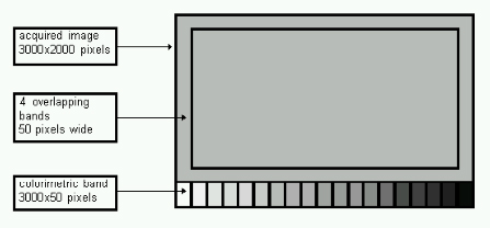
Two interesting points to be developed regard reproducibility of optical conditions during image acquisition and uncertainty evaluation of acquired digital values. A surface moving with the camera, having an area of about 3% of the whole image (3000x2000), is used for colorimetric analysis (see Figure 5). The surface, rigidly connected to the camera by means of an arm, is subdivided into several zones, each characterized by a particular known color. Squares having known chromatic characteristics such as pure copper, gold and silver, previously calibrated by a spectrometer, may be included.
 |
The Shroud may be positioned vertically in a room. A distance of at least 5 m is needed in front of the Shroud for proper acquisition of images. The apparatus, to be carefully studied after the choice of the most appropriate CCD sensor, with relative camera, preliminarily consists of:
- previously calibrated RGB camera, with a spatial resolution of about 3000x2000 square pixels (12-bit gray level, 24x36mm).
- a band for colorimetry attached to the camera.
- stiff tripod with 3 wheels to allow translation along a slide-bar on the floor.
- at least 4 halogen lamps mounted on the tripod.
- various fixed-focus macro lenses; a bellows will also be used.
- personal computer.
5) UNCERTAINTY ANALYSIS
Development of uncertainty analysis is necessary before, during and after Shroud mapping if qualified data are acquired.
a) uncertainty analysis before mapping to calibrate the whole system.
In this phase uncertainty sources are considered without distinction between Type A and Type B (ISO Guide [13]), and all sources of disturbances are presumed to be controlled. Evaluation of uncertainty in the Project Phase may be obtained by combining the modeling uncertainty of the whole acquisition system, the resolution uncertainty of each sensor in the measuring chain, and instrumental uncertainty (see Figure 6). In particular, the following aspects must be investigated:
- A) Spatial and chromatic resolution.
- B) Temporal and spatial stability.
- C) Distortions of the optical system. Low distortion lenses are needed. The camera with each lens will be spatially calibrated in order to evaluate electro-optical distortion effects and to detect effective pixel side ratio.
- D) Dark current effects signal-to-noise ratio, position of defective pixels, clusters, etc.
 |
b) Uncertainty analysis during mapping.
the following aspects must be investigated:
- A) Temporal and spatial stability of both acquisition system and light source
- B) Possible noise sources, such as support vibrations, temperature variations, etc.
- C) Repeatability and reproducibility of acquired data.
c) uncertainty analysis after mapping.
the procedure consists of the following steps:
- 1) evaluation of uncertainty components due to system modeling, calibration, data acquisition and data reduction;
- 2) evaluation of bias and repeatability of acquire data;
- 3) propagation of uncertainty to the results (spatial and chromatic data of each image) must be evaluated in terms of Type A and Type B components (ISO Guide[13]), evaluating all effects such as repeatability, reproducibility, systematic effects, etc.
taking into account for chromatic calibration (controlled a posteriori by direct comparison of acquired RGB values corresponding to the reference surface of each image). Relative uncertainty values will be associated with all the chromatic and spatial data of all acquired images.
6) CONCLUSIONS
High resolution colorimetric mapping of the Turin Shroud, by means of digital acquisition with a solid state camera, is proposed, with the aim of obtaining a data base for future accurate analyses. For example, more information about the mechanism of the image formation on the cloth may be obtained.
In this work, some preliminary problems, regarding choice of the experimental apparatus and analysis of metrological problems, connected to the acquisition system are discussed. In particular, proper calibration of the acquisition systems with uncertainty evaluation (done before, during and after mapping) considering some metrological problems such as the spatial and temporal stability of acquired signals, image distortions, dark currents and defective pixel effects are foreseen to maximize the quality of acquired data.
REFERENCES
1) Emanuela Marinelli: "La Sindone, un'immagine 'impossibile' ", ed. San Paolo, Alba (Cuneo), Italy, 1996.
2) Gino Moretto: "Sindone-la guida", editrice Elle Di Ci, Leumann (Torino), Italy,1996.
3) Eric J. Jumper, Robert W Mottern: "Scientific investigation of the Shroud of Turin", Applied Optics, 15 June 1980, Vol. 19, n°12.
4) John H. Heller, Alan D. Adler: " Blood on the Shroud of Turin", Applied Optics, 15 August 1980, Vol. 19, n°16
5) Roger Gilbert Jr., Marion M. Gilbert: " Ultraviolet-visible reflactance and fluorescence spectra of thew Shroud of Turin", Applied Optics, 15 June 1980, Vol. 19, n°12.
6) S.F. Pellicori: "Spectral properties of the Shroud of Turin", Applied Optiics, 15 June 1980, Vol. 19, n°12.
7) John P: Jackson, Eric J. Jumper, William R: Ercoline: " Correlation of image intensity on the Turin Shroud with the 3-D structure of a human body shape", Applied Optiics, 15 July 1984, Vol. 23, n°14.
8) P.E. Damon et al.: "Radiocarbon dating of the Shroud of Turin", Nature, vol. 337, 16 February 1989, pagg. 611-615.
9) H.E. Gove, S.J. Mattingly, A.R. David, L.A. Garza-Valdes: A problematic source of organic contamination of linen, Nuclear Instruments and Methods in Physics Research, B 123 (1997) 504-507.
10) E. Garello, M: Salomoni: "Datazione della Sindone attraverso la luminescenza all'infrarosso", atti del IV Congresso Nazionale di Siracusa, ed. Paoline, Cinisello B., (Milano), 1988.
11) Philips: "FTF3020M: Preliminary Data Sheet, Philips Electronics N.V., nÝ9922 157 31001/190-1, 1996.
12) Thierry Aubry, Jim Belsky "Selection of Solid-State Detectors", EuroPhotonich, December/January 1997.
13) "Guide to the Expression of the Uncertainty in Measurement", ISO, 1993.